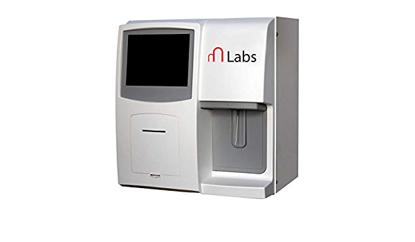
Hematology analyzers (sometimes spelled hematology analyzers in British English) are used to quickly and accurately count and identify blood cells. Laboratory staff counted each solitary blood cell under a microscope in the 1950s. This was succeeded by Wallace H. Coulter’s first, extremely rudimentary hematology analyzer, which was tedious and inconsistent. Coulter’s Principle was used by the first hematology analyzers (see Coulter counter). They have, however, grown to include a wide range of procedures.
Techniques of Hematology Analyzer:
- Electrical impedance
The Coulter Principle, often known as electrical impedance, is a standard method for counting cells. It’s used by almost every hematological analyzer.
- Flow cytometry
Laser flow cytometry is more expensive than impedance analysis due to the requirement for expensive chemicals, but it offers more information on the morphology of blood cells. It’s an excellent method for calculating five-part WBC differentials.
- Fluorescent flow cytometry
Thanks to the inclusion of fluorescent reagents, flow cytometry may now be used to analyze individual cell populations. Fluorescent colors indicate the nucleus-plasma ratio of each tagged cell. It may be used to examine platelets, nucleated RBCs, and reticulocytes. Cellular fluorescence is used to assess RNA (reticulocytes), DNA (nucleated red cells), and cell surface antigens.
- Light scatter
In a single line, each cell travels through a flow cell. When a laser device is pointed towards a flow cell, the laser light beam is distributed in a variety of directions. A forward scatter light detector (also known as a FALS detector) gathers light that is proportional to the size of the cell.
- Light absorption
Absorption spectrophotometry is used to quantify the concentration of hemoglobin after it has been transformed to cyanmethemoglobin or another molecule. Certain analyzers use peroxidase cytochemistry to classify leukocytes; peroxidase activity is measured by absorbance.
- Electrical conductivity
To identify leucocytes based on their physical and chemical makeup, several analyzers use the conductivity of high-frequency electricity.
Types of Hematology Analyzer:
- 3-Part Differential Cell Counter
A three-part differential cell counter is used to determine the size and volume of a cell using Coulter’s Principle. The sample is lysed and dissolved in an electrolyte solution in a container that also houses a smaller container. Two pumps transport the solution from the larger container to the smaller container, one creating a vacuum and the other replenishing the lost solution. The smaller container has a small aperture (an orifice) towards the bottom. Coulter’s proposal is implemented using two electrodes.
- 5-Part Differential Cell Counter
This type of hematology analyzer uses both Coulter’s Principle and flow cytometry to determine the granularity, diameter, and inner complexity of the cells. Using hydrodynamic focusing, the cells are passed through an aperture one at a time. During this procedure, a laser is pointed at them, and the scattered light is measured from various angles. Absorbance is also taken into consideration.
Uses of Hematology Analyzer:
Hematology analyzers are used to produce a complete blood count (CBC), which is typically the first test requested by doctors to determine a patient’s general health status. A complete blood count includes red blood cell (RBC), white blood cell (WBC), hemoglobin, and platelet counts, as well as hematocrit values.
- The breadth of the RBC distribution
- The volume of the average corpuscle
- Hemoglobin concentration in the blood
- Concentrations of mean corpuscular hemoglobin
- The percentage and the absolute value of the WBC differential count
- The breadth of the platelet dispersion
- The average amount of platelets
- The platelet cell ratio is high.
- Criteria for platelets
What is a Hematology Stain?
Hematology stains and reagents are required for clinical and medical cytology research. Blood, bone marrow, and lymphatic system components that are difficult to detect are stained and identified. On prepared blood smears, whether manually or machine processed, the Wright, Giesma, and Leishman formulas work well.
Components of hematologic Stain:
- Routine hematologic stains:
Romanowsky type dyes, such as a modified Wright’s stain (used at Cornell University’s Clinical Pathology Laboratory), and rapid stains, such as Diff-quik®, are used to stain blood smears. These stains employ both acidic (red) and basic (blue) dyes. In smears, nuclei and RNA in red blood cells (RBCs) appear blue, whereas hemoglobin in RBCs appears red.
- Reticulocytes:
Reticulocytes are immature anucleate RBCs whose cytoplasmic RNA has been stained with a fluorescent dye (e.g. thiazole orange) or precipitated with a supravital dye (e.g. new methylene blue) for laser detection. At Cornell, reticulocytes are identified and counted by their ability to absorb Oxazine-750, a nucleic acid-binding dye.
Conclusion
Hematology analyzers are specialized machines that count the number of red and white blood cells, blood platelets, hemoglobin, and haematocrit in a sample of blood. The results of various blood tests can be used to detect a variety of hematologic disorders. Blood analysis can reveal anemia, diabetes, autoimmune deficiencies, infections, and cancers. It is possible to discover genetic information, illnesses, and organ shortages.


